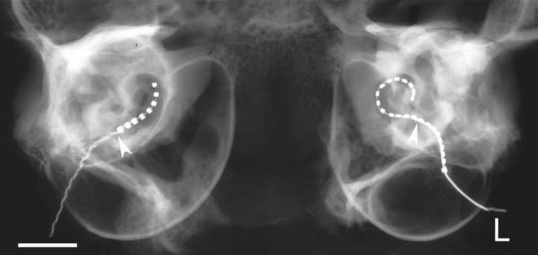
Figure 2.
A micro-focus radiograph of a standard 8 ring electrode array (right cochlea) and a Hybrid-L24 array (L; left cochlea) illustrating the typical insertion profiles obtained with each array in the present study. Both arrays were inserted via the round window to the point of first resistance. The arrowhead illustrates the site of the round window. Scale bar = 5 mm.

