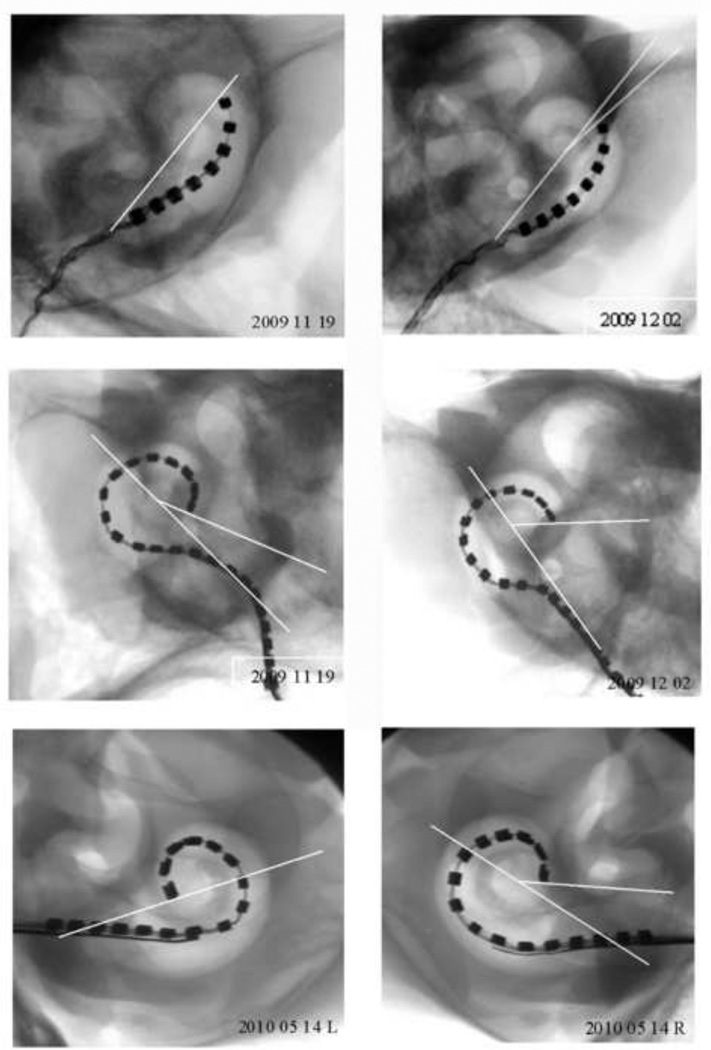
Figure 3.
Micro-focus radiographs from all six cochleae used in this study (two standard, two Hybrid-L24 and two Hybrid-L16 electrode arrays). The insertion depth (in degrees) were measured to the tip of each array by drawing a 180° reference line from the middle of the round window to the centre point of the cochlear spiral then creating a second angle from the centre point to the tip of the array. Scale bar = 5 mm.

