
Figure 6.
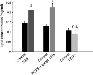
Membrane fragmentation induced by prolonged incubation with Aβ1-40. Lipid concentrations in the supernatant after centrifugation from brain extract LUVs (dark gray bar) and POPC/POPS/gangliosides 5.5/3/1.5 LUVs (gray bar) after incubation with Aβ1-40 for 48 h are shown. The failure of lipid vesicles to sediment is an indication of their disruption to smaller micelle-like structures. No significant lipids were detected in the supernatant of samples containing POPC/POPS 7:3 (light gray bar), in agreement with the 6-carboxyfluorescein dye leakage assay (Fig. 1). Results are the average of three independent measures, and error bars represent the standard deviation.