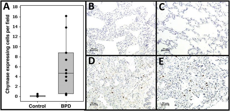
Figure 6.
Increased connective tissue–type mast cell accumulation in bronchopulmonary dysplasia (BPD) lungs. We used immunohistochemistry to identify chymase-expressing cells, a specific marker for connective tissue–type mast cells. (A) Quantitative analysis of the number of chymase-expressing cells in BPD and non-BPD control lungs. Shown are the average numbers of stained cells/field for each individual subject (dots), the group means (bar), and interquartile range (box). (B, C) Examples of staining patterns in age-matched control subjects. (D, E) Examples of staining patterns in BPD lungs. Magnification is as indicated on scale bars. A significant approximately 50-fold increase in chymase-positive cells is observed in BPD lungs.

