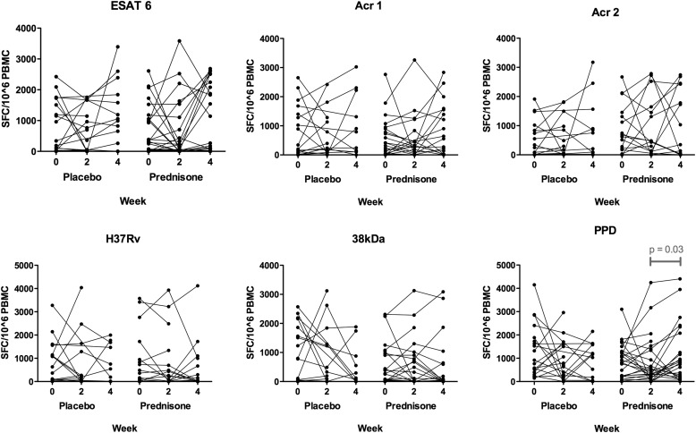
Figure 1.
Enzyme-linked immunospot results in response to overnight restimulation of peripheral blood mononuclear cells (PBMCs) with six different antigen stimuli at 0, 2, and 4 weeks, comparing placebo- and prednisone-treated participants. Antigen stimuli used were as follows: early secreted antigen target-6 (ESAT-6, 5 μg/ml), α-crystallin 1 (Acr1; 10 μg/ml), α-crystallin 2 (Acr2; 20 μg/ml), and 38-kD antigen (2.5 μg/ml); purified protein derivative (PPD) was used at 200 IU/ml (4.6 μg/ml) and heat-killed H37Rv Mycobacterium tuberculosis was used at a multiplicity of infection of 1:1 (200,000 H37Rv:200,000 PBMCs). Four patients in the placebo-treated arm switched to open-label prednisone at Week 2 and thus their Week 4 samples were excluded from analysis. Statistical comparisons of median values for each time point comparing placebo- and prednisone-treated participants showed no significant differences. Within-group paired comparisons between time points (0 vs. 2, 0 vs. 4, and 2 vs. 4 wk) revealed only one significant difference (P < 0.05): a rise in the number of spot-forming cells (SFCs) to PPD in the prednisone-treated participants from 2 to 4 weeks (from 449 SFCs/106 PBMCs [IQR, 197–1,368] at Week 2 to 800 [IQR, 188–2,078] at Week 4 [P = 0.03]).

