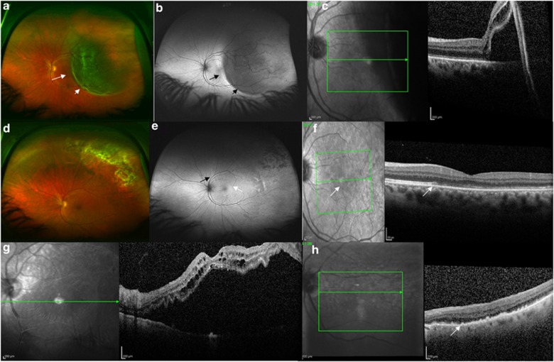
Figure 2.
(a) Preoperative wide-field colour image of a 50-year-old female patient with a RRD involving the fovea. The short arrow identifies the area of bullous retinal detachment readily identified with ophthalmoscopy. The long arrow identifies an area of shallow neurosensory detachment extending through the fovea. (b) Preoperative wide field AF image of the same patient demonstrating the area of bullous detachment as hypofluorescence (short arrow) and the HLE with the long black arrow, indicating the area of shallow neurosensory detachment. (c) Preoperative OCT image of the patient from panels (a) and (b). The boundary between detached and attached retina is readily identified. (d) One-month postoperative colour image demonstrating reattachment of the retina with laser chorioretinal scars in the superotemporal quadrant. (e) One-month peripheral AF image demonstrating the residual demarcation line (black arrow) and granular AF changes (white arrow) within the area of previously detached retina. (f) Postoperative OCT image of the patient. A white arrow on the infrared image highlights the boundary between attached and detached retina, preoperatively. The OCT shows disruption of the IS/OS junction in the region of previously detached retina (which showed granular changes with AF) and a white arrow shows the boundary. (g) Preoperative OCT image of a different patient (78-year-old male) with a macula-affecting retinal detachment. (h) Postoperative OCT image of the patient from panel (g), who demonstrated postoperative granular AF changes, demonstrating significant outer retinal disturbance. The outer retinal changes of the IS/OS junction appear similar to the subretinal or outer retinal deposits (white arrow).

