
Figure 3. PTEN inhibited CXCR4-mediated migration and proliferation.
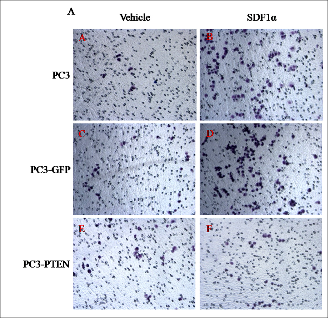


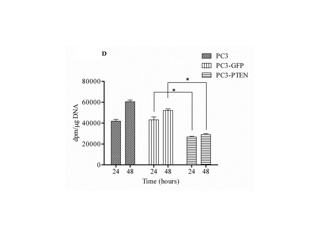
(A) Cells were transiently transfected with 4 µg of pcDNA3-GFP or pcDNA3-GFP-PTEN constructs, prior to serum-starvation for 24 h. Cells (2×10⁴) were added to the upper transwell chamber and allowed to migrate towards 100 ng/mL of SDF1α in the lower wells for 4 h at 37°C. Five fields of each transwell insert were randomly selected and counted for migrated cells at 10X magnification. Micrographs of migrated cells were taken at 10X magnification using a Zeiss Axiovert 200M light microscope. (B) A graphical representation of total migrated cells. Experiments were repeated thrice and data represents the averages of three independent experiments. (C) Cells were transiently transfected and serum starved, prior to treatment with 100ng/mL of SDF1α at the indicated time points. MTT assays were performed after each time point following the manufacturer’s protocol. Experiments were repeated thrice in triplicate and data represents the averages of three independent experiments. (D) Cells were transiently transfected and serum starved, prior to treatment with 100ng/mL of SDF1α at the indicated time points. Proliferation was measured by [3H] thymidine uptake/DNA synthesis assay. (*, P < 0.05).