
Figure 4. PTEN inhibited CXCR4-mediated migration by inhibiting ERK1/2 phosphorylation.
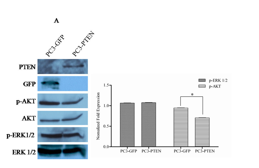
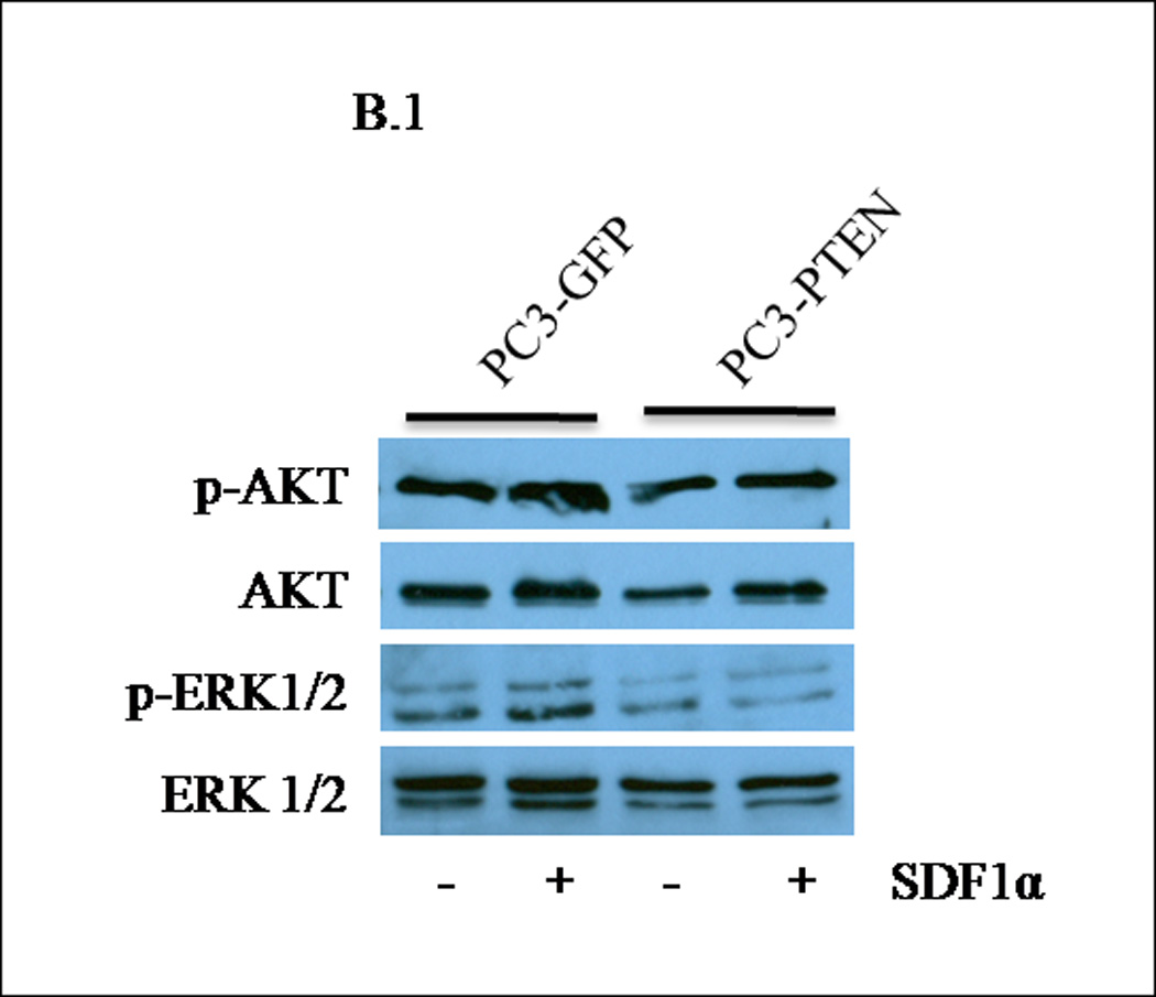
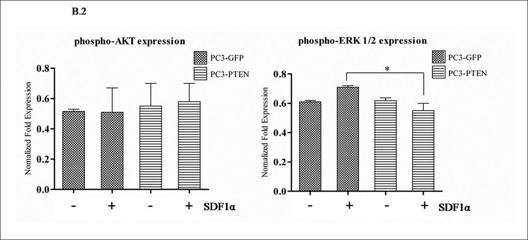
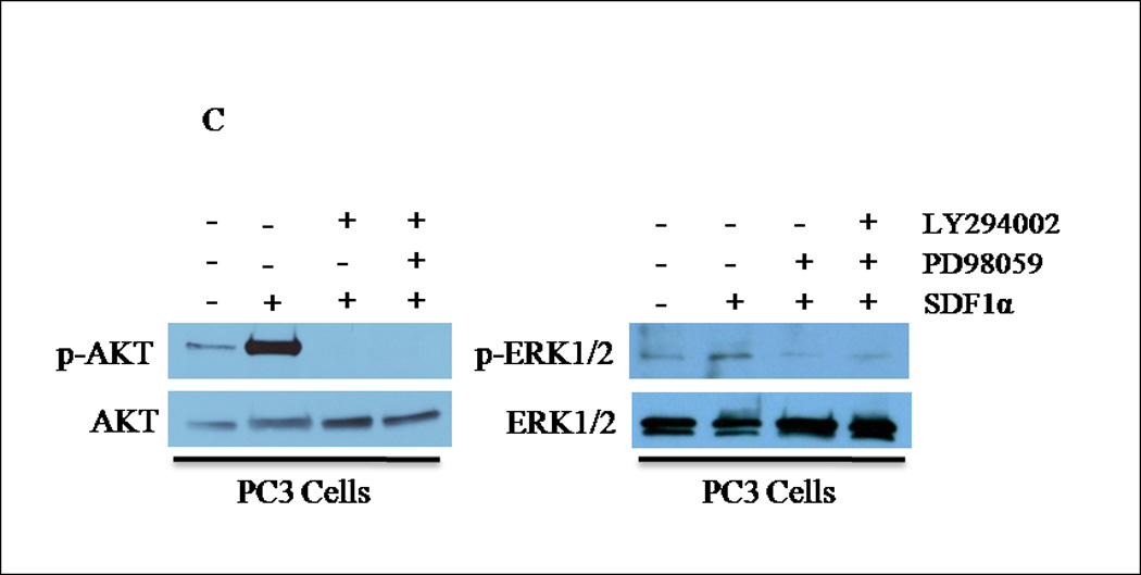
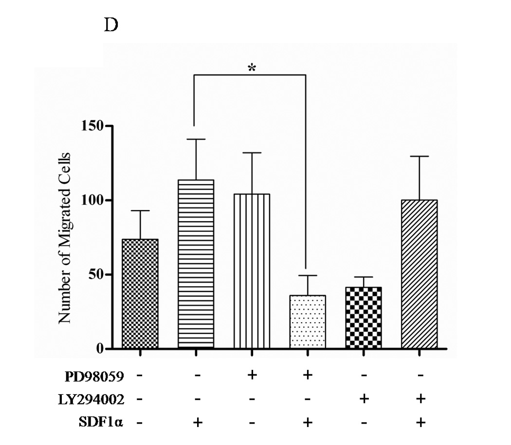
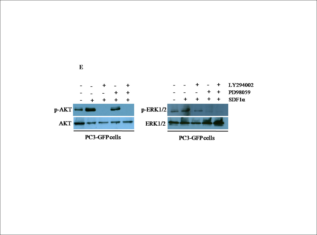

(A) Transiently transfected cells were lysed and 30 µg of protein were analyzed for PTEN, p-AKT, total AKT, p-ERK1/2 and total ERK1/2 expression by western blot analysis using specific antibodies. Graph represents a densitometric analysis for the relative expression of p-AKT and p-ERK1/2 compared to controls. (B) Transiently transfected cells were treated with 100 ng/mL of SDF1α for 10 minutes followed by western blot analysis of 30 µg of protein using p-AKT, total AKT, p-ERK1/2 and total ERK1/2 specific antibodies. Graph represents a densitometric analysis for the relative expression of p-AKT and p-ERK1/2 compared to controls. (C) PC3 and (E) PC3-GFP cells were serum starved prior to treatment with 50mM of PD98059 and/or 10mM of LY294002 for 1 h at 37°C. Cells were then stimulated with 100 ng/mL of SDF1α for 10 minutes followed by western blot analysis of 30 µg of protein using p-AKT, total AKT, p-ERK and total ERK1/2 specific antibodies. (D) PC3 and (F) PC3-GFP cells were serum starved and treated with 50 µM of PD98059 and/or 10 µM of LY294002 for 1 h at 37°C. Cells (2×10⁴) were added to the upper transwell chamber and allowed to migrate towards 100 ng/mL of SDF1α in the lower wells for 6 h at 37°C. Five fields were randomly selected and counted for migrated cells at 10X magnification. Experiments were repeated thrice and data represents the averages of three independent experiments. (*, P < 0.05).