
Figure 3.
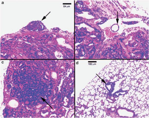
Hematoxylin and eosin (H&E) staining of thyroids and lungs of representative sections from TRα1−/− TRβ−/− mice. Histological sections from tissues of mice showed evidence of capsular invasion in thyroid (a) (arrow), vascular invasion in thyroid (b) (arrow), anaplasia in thyroid (c) and metastatic thyroid carcinoma lesions in lung (d) (arrow).