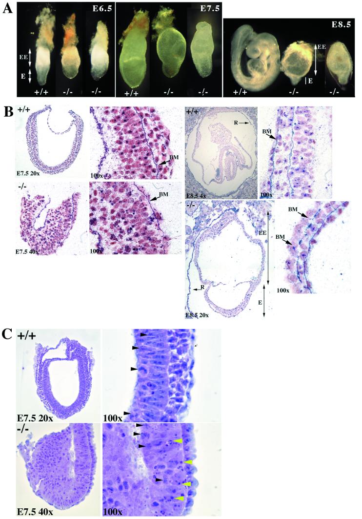
Figure 2.
Phenotypic analysis of wild-type and mutant embryos. (A) Whole-mount gross morphology of wild-type and Ctr1−/− embryos at E6.5, E7.5, and E8.5. The embryos are oriented such that the embryonic region (E) is located at the bottom and the extra-embryonic structures are on the top (EE). All embryos were genotyped by PCR after photography. Identical phenotypes were observed in both the 124 and 191 lines, and wild-type embryos were indistinguishable from heterozygotes. Photographs were taken on a Nikon SMZ 800 dissecting microscope. E6.5 embryos were photographed at ×6.5, E7.5 at ×5, and E8.5 at ×4 with a ×2.5 projection lens. (B) Histological examination of sectioned embryos. E7.5 and E8.5 wild-type and mutant were stained with anti-collagen IV staining and counterstained with Fast Red. At E7.5, the basement membrane (BM) of the mutant embryo appears intact. At E8.5, the mutant extra-embryonic region (EE) and underdeveloped embryonic region (E) are seen. Note the Reichert's membrane (R) is broken in the E8.5 mutants but intact in the wild-type E8.5. At ×100, the basement membrane is intact in the wild type, but gaps along the basement membrane are apparent in the mutant, as indicated by the arrows. (C) Hematoxylin and eosin staining of sagittal sections of E7.5 wild-type and mutant embryos. Only the embryonic region is shown because the upper extra-embryonic region was removed for genotyping by PCR. At ×100 in the mutant, areas of apoptosis can be seen (indicated by yellow arrowheads); however, mitosis is unaffected (indicated by black arrowheads). In the wild-type normal section at ×100, mitosis can be seen as indicated by the black arrowheads.

