Figure 3.
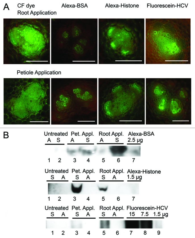
Analysis of symplastic and apoplastic accumulation of fluorescent dyes and proteins in B. oleracea leaves. (A) Confocal microscopic images of CF dye, Alexa-BSA and Alexa-Histone H1 in fan-shaped bundles in petiole cross sections following root application or L1 petiole application. Bar = 50μm. CF dye fills the vascular bundle and spreads to surrounding tissues. Alexa-BSA was mainly in the tracheary elements of the xylem. Alexa-Histone spreads beyond the phloem into parenchyma cells. Fluorescence-HCV spreads further than Alexa-BSA but not as far as Alexa-Histone throughout the vascular bundle. Control samples were treated with buffer and show no fluorescence (not shown here). (B) Immunoblot probed with polyclonal antisera detecting BSA and Histone. Pet. Appl, petiole application; Root Appl, root application; M, marker; Plant endogenous histone H1 is not also detected by antisera. Apoplastic wash fluids (A) and cell extracts (S) were pooled from L3 and L4 leaves. The top immunoblot detects Alexa-BSA in both the A and S extracts. Alexa-BSA was directly loaded to gel and produces a band around 66 kDa (lane 7). The middle immunoblot detects Alexa-Histone in the S but not A wash fluid. Alexa-Histone was directly loaded to the gel and produces a band that is around 34 kD. The bottom immunoblot detects fluorescein-HCV in the S but not A fraction. Lanes 7–9 contain samples of fluorescein-HCV that were directly loaded to the gel. Loading amounts (μg) for Alexa-BSA and -Histone and fluorescein-HCV are indicated above the lanes.

