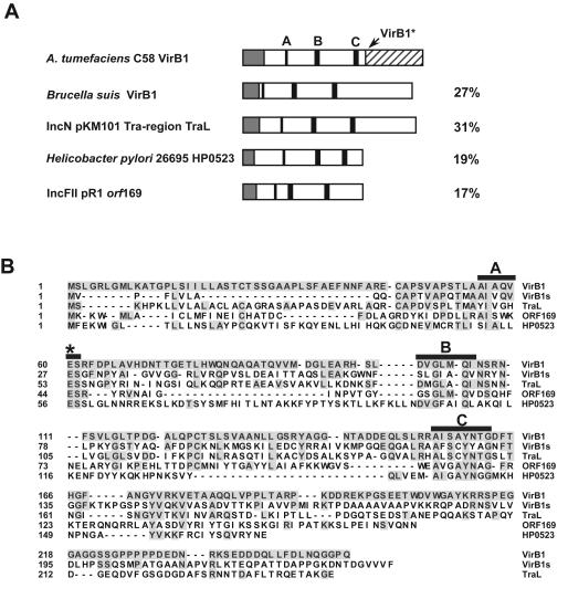
FIG. 1.
VirB1 orthologs and active-site mutants used in this study. (A) Schematic representation of the analyzed proteins and their amino acid sequence identities (percentages noted on the right) to A. tumefaciens C58 VirB1. Amino acid residues in boxes A, B, and C (black boxes) are implicated in the enzymatic activity of lytic transglycosylases. They were assigned in the different orthologs according to the method of Bayer et al. (5). Signal peptides (grey boxes) were assigned by the SignalP V1.1 algorithm (http://www.cbs.dtu.dk/services/SignalP). The C-terminal processing site of VirB1 (A173QQ) is shown as a hatched box. (B) Alignment of VirB1 orthologs investigated in this study; residues identical to those of VirB1 are shaded. Conserved residues are indicated by black boxes A, B, and C, and the putative active-site Glu residues are labeled with a star. Active-site residues corresponding to Glu60 from A. tumefaciens VirB1 were changed to Ala in VirB1s and TraL in the course of this study. The alignment was generated with the MegAlign program (DNA Star).

