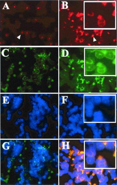
Figure 2.
Immunofluorescence to detect cell type and subcellular localization. Bone marrow cells from transgenic mouse (B, D, F, and H) and the nontransgenic littermate control (A, C, E, and G) were prepared by using a cytospin apparatus, and incubated simultaneously with antibody against cyclin A1 (A and B) and antibody against antigen 7/4 (C and D), as well as DAPI (4′,6-diamidino-2-phenylindole; E and F). Red blood cells also showed signals due to their nonspecific cross-reaction with dyes (indicated by white arrowhead in A and B). Note that cyclin A1 antibody strongly stained with the cells with myeloid precursors (B). The colocalization of cyclin A1 and 7/4 was detected in myeloid precursors of the transgenic line (H). Although cells with a neutrophilic morphology weakly expressed cyclin A1 (red staining in cells at top of Inset in B), the green staining for antigen 7/4 was significantly more intense (Inset in D). These data suggested that although both cyclin A1 and antigen 7/4 are expressed in myeloid cells, cyclin A1 appears to be more abundant in earlier stages of differentiation than in later stages. It is also noted that we did not detect cyclin A1 protein in myeloid cells in the nontransgenic littermate BM (A and G).

