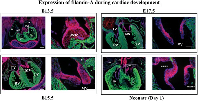
Figure 1.
Protein expression of filamin-A during cardiogenesis. Immunohistochemistry (IHC) was performed on E13.5, E15.5, E17.5, and neonatal Day 1 hearts for filamin-A (red) and MF20 (myocytes-green). Each timepoint shows a low magnification and higher magnification of boxed region, primarily focusing on the development of the mitral valve leaflets. Filamin-A is robustly expressed throughout cardiac development, being expressed in interstitial cells of the atrioventricular septal complex (AVSC), the mitral and tricuspid valves (MV and TV), the epicardium, AV sulcus (arrow), endocardial cells, developing blood vessels including coronaries and aortae (Ao). Expression is not detected in cardiomyocytes. Scale bars: low magnification = 200 µm, high magnification = 100 µm. RV, right ventricle; LV, left ventricle; RA, right atrium; LA, left atrium; AoLMiV, aortic leaflet of the mitral valve; MuLMiV, mural leaflet of the mitral valve; iAVC, inferior AV cushion; rAVC, right AV cushion.

