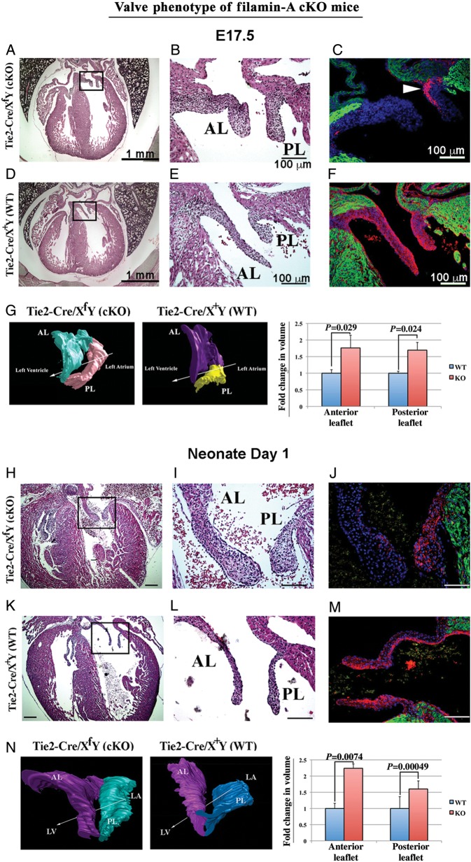
Figure 3.
Filamin-A cKO mice exhibit enlarged AV leaflets during foetal and neonatal life. (A, B, D, E, H, I, K, and L) The histological assessment of filamin-A conditional KO mice (cKO; Tie2-Cre/XfY) compared with littermate control animals (Tie2-Cre/X+Y) at E17.5 and neonatal Day 2 using H&E stains. Mitral leaflets of the cKO mice exhibit significant changes in the valve width, length, and shape. (C, F, J, and M) IHC to confirm filamin-A is genetically removed using the Tie2-Cre recombinase mouse line. Little staining of filamin-A (red) is seen in the anterior leaflet of the cKO mouse (AL), whereas significant filamin-A positivity is observed in the posterior leaflet (PL, arrowhead). Green staining is MF20. (G and N) AMIRA 3D reconstructions were performed on the entire mitral leaflet and show shape modifications in the cKO coincident with a significant increase (P-values noted) in volume. Arrows in G and N indicate direction of blood flow. n = 3 for each genotype. All samples were littermates, sex, and age matched. Magnification bars: A, D, H, K = 1 mm; B, C, E, F, I, J, L, M = 100 μm.

