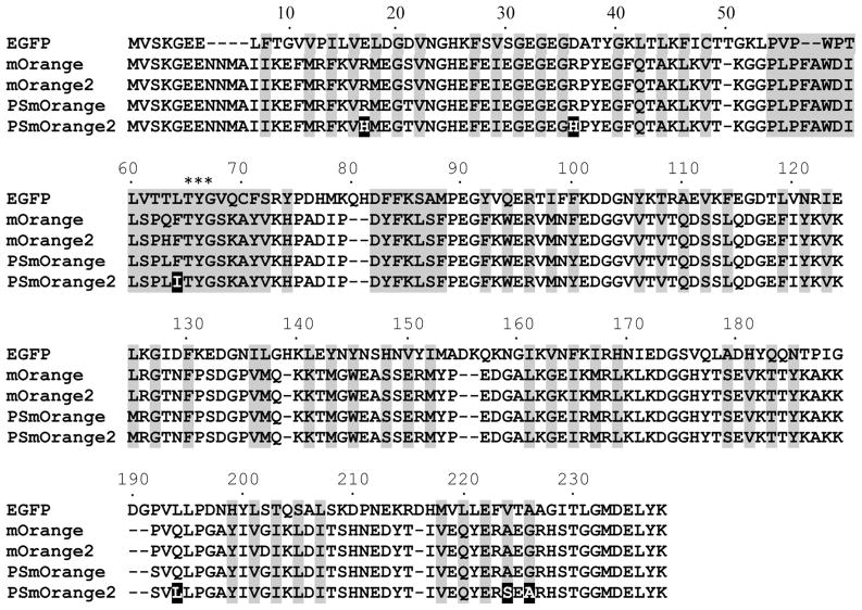
Figure 1.
Alignment of the amino acid sequences for the EGFP, mOrange, mOrange2, PSmOrange and PSmOrange2 proteins. Alignment numbering follows that of EGFP. Residues buried in the protein β-barrel fold are shaded. Asterisks indicate residues that form the chromophore. Mutations in PSmOrange relative to PSmOrange are indicated in white on black background.

