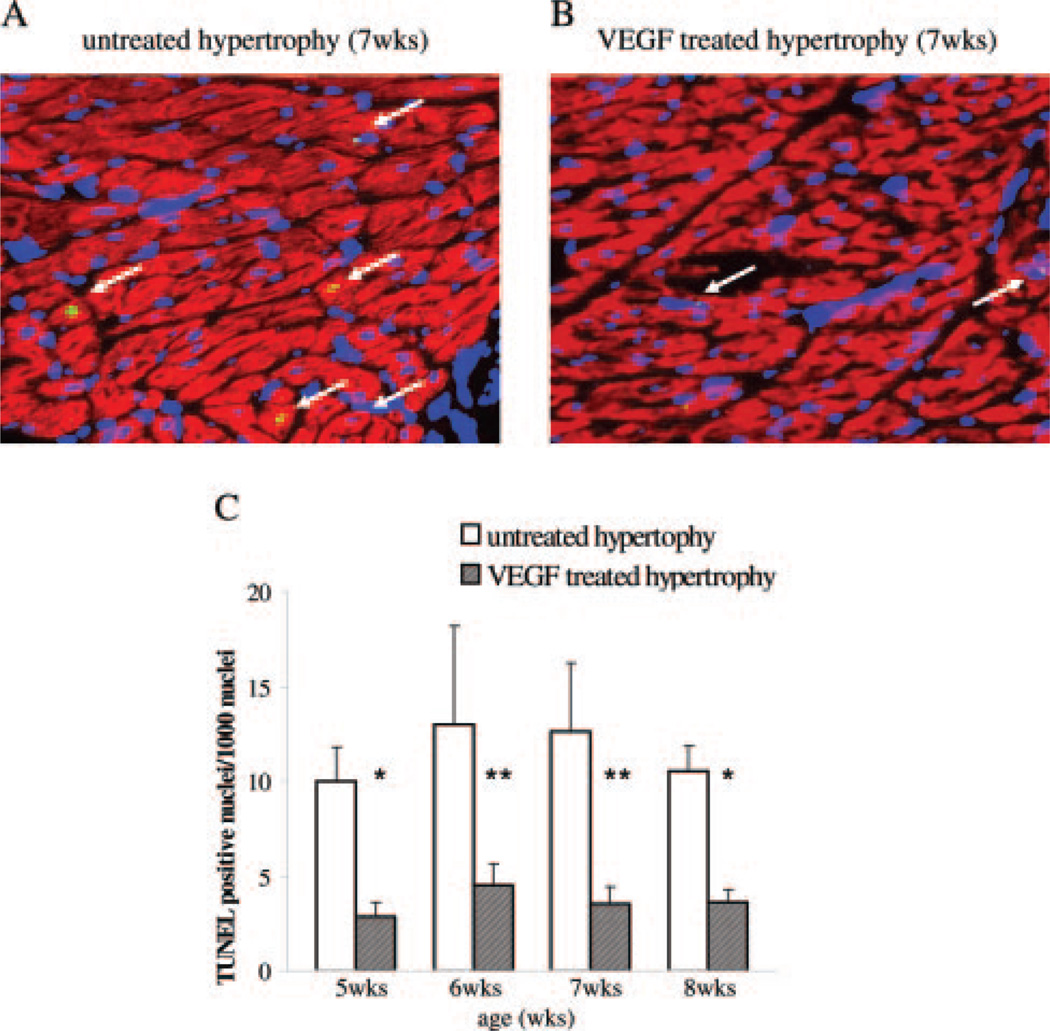
Figure 2.
TUNEL staining of untreated and VEGF-treated hypertrophy. A and B, Representative immunohistochemical sections showing TUNEL staining are shown. Apoptotic nuclei (arrow) are shown by green fluorescence and the localization of nuclei is documented by DAPI counterstaining and labeling of cardiomyocytes with red fluorescent desmin. Hearts at 7 weeks of age from untreated hypertrophied animals and 1 week after the second VEGF administration are depicted. All sections are shown at the same magnification. C, The summarized results are expressed as TUNEL positive cardiomyocyte nuclei per 1000 nuclei. VEGF treatment prevented apoptotic cell death in progressive hypertrophy. *P<0.05 and **P<0.01 vs untreated hypertrophy.

