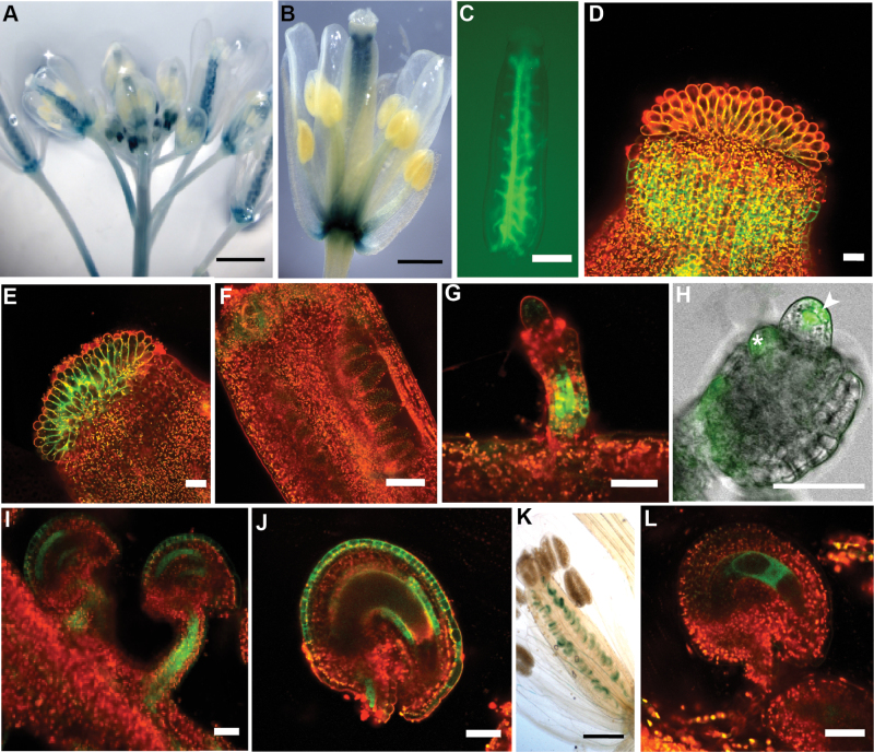
Fig. 1.
FLP and MYB88 are expressed in reproductive organs. (A–J) Micrographs of pFLP::GUS-GFP transgenic plants. (A and B) GUS staining. (C–J) GFP fluorescence visualized using confocal microscopy. (A) FLP expression in whole inflorescence. (B) FLP is expressed in the carpel. (C) Expression of FLP can be seen in the placenta. (D) FLP is expressed in the style. (E) FLP is expressed in the stigmatic tissue. (F) During early ovule development, before integument initiation, little FLP expression is detectable within the ovule. (G) Expression of FLP can be seen in the funiculus as the integuments are initiating. (H) FLP is expressed in the nucellus, including epidermal cells (arrowhead) and the MMC. FLP is also expressed in initiating integuments (asterisk) and the funiculus. (I) FLP expression persists in the funiculus and is seen in the integuments of older ovules. (J) In stage 13 ovules, FLP is expressed in both the endothelial layer and the outer layer of the outer integument. (K and L) Micrographs of pMYB88::GUS-GFP transgenic plants. (K) GUS staining showing MYB88 expression in ovules. (L) In stage 13 ovules, MYB88 is expressed in the embryo sac.

