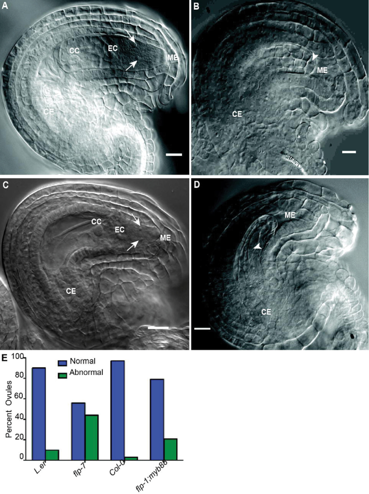
Fig. 3.
Loss of FLP and/or MYB88 leads to abnormal nucellar structures. (A–D) DIC micrographs of FG7 ovules containing mature female gametophytes. (A) Col-0. (B) flp-1; myb88. (C) L. er. (D) flp-7. (E) Quantification of embryo sac defects. CC, central cell; CE, chalazal end of the embryo sac; EC, egg cell; ME, micropylar end of the embryo sac. Arrows indicate synergid cells, and arrowheads indicate large cells found in abnormal flp ovules in the region where an embryo sac would normally form.

