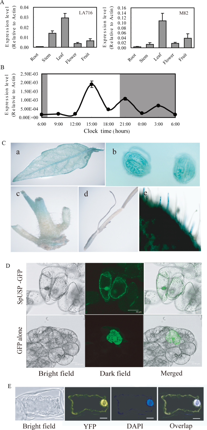
Fig. 1.
Expression patterns of SpUSP. (A) Tissue profiling analysis of SpUSP in different organs of wild tomato LA716 (Solanum pennellii) and cultivated tomato M82 (S. lycopersicum) using qRT-PCR. (B) Expression pattern of SpUSP during a 24h period. Leaf samples were collected every 3h for 24h starting from 06.00h. (C) Expression patterns of SpUSP via GUS staining: (a) leaf, (b) stoma, (c) stem, (d) root, and (e) trichome. (D) Subcellular localization of SpUSP. The photographs were taken under bright light, in the dark field for the GFP-derived green fluorescence and merged respectively. (E) Interaction of SpUSP with annexin via BiFC. The photographs were taken under bright light, in the dark field for YFP-derived green fluorescence, staining with DAPI and overlap, respectively. Scale bars=10 μm.

