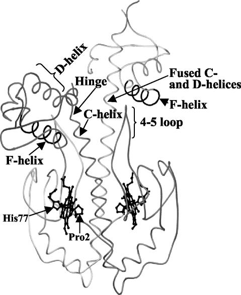
FIG. 1.
Inactive Fe(II) CooA structure adapted from that of the strain with PDB identification no. 1FT9. The protein consists of two monomers, shaded differently in this figure, which dimerize along the central C-helices of adjacent effector-binding domains. The solved structure is asymmetric, in which one monomer contains fused C- and D-helices (20). Nonetheless, both F-helices that interact with DNA in a sequence-specific manner are buried from the surface in the structure. The 4/5 loop is noted and so are the Pro2 and His77 heme Fe(II) ligands. In these representations, the DNA-binding domains form the upper part of each structure and the positions of the helices that specifically interact with DNA are designated as the F-helix.

