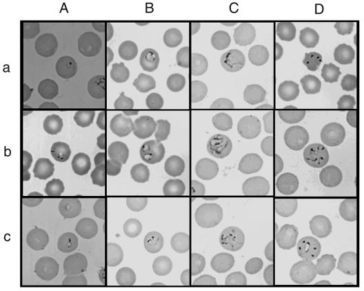
FIG. 1.
Photomicrographs of the Thai babesia on Giemsa-stained thin blood smear of babesia-positive Bandicota indica. (A) Panel a, a ring-shaped trophozoite consisting of a cytoplasmic rim with a chromatin dot; panel b, double infection of ring-shaped trophozoites with double chromatin dots and a vacuolated lesion on the cytoplasmic rim; panel c, a pyriform-shaped trophozoite. (B) Panel a, a large ring or annular form with three chromatin masses, two dot-like and one elongated; panel b, double infection of annular trophozoites; panel c, a trophozoite with bridge-like cytoplasms. (C) Panel a, an irregular large trophozoite; panel b, double infection of irregular large trophozoites; panel c, an irregular form with multiple chromatin dots. (D) Panel a, Maltese-cross-like form; panel b, four trophozoites, presumably developing independently; panel c, five trophozoites observed in one red blood cell.

