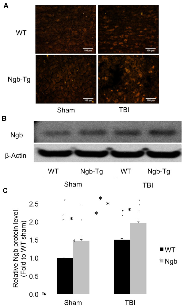
Figure 1 .
Ngb expression levels in mouse brains after TBI.A. Representative immunohistochemistry of Ngb protein expression in WT and Ngb-Tg mice cortex of peri-lesion area at sham animals and at 6 h after TBI. Original magnification × 200 in all photomicrographs. B. Representative western blot of Ngb protein expression in WT and Ngb-Tg mice cortex of peri-lesion area at sham animals and at 6 h after TBI, β-actin served as equal protein loading controls. C. Quantitation of Ngb protein levels. Data were expressed as mean ± SD. *P < 0.05, n = 5 per group.

