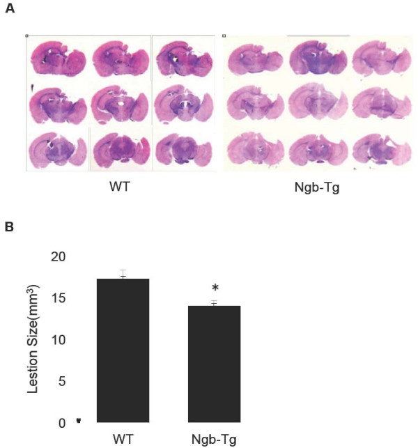
Figure 4 .
Measurements of cortical lesion volume in WT and Ngb-Tg mice.A. Representative photomicrographs of the traumatic lesions in H&E stained WT and Ngb-Tg mouse brain sections at 21 days after TBI. B. Traumatic brain lesion size. Data were expressed as mean ± SD. *P < 0.05, n = 15 for WT and 11 for Ngb-Tg per group, respectively.

