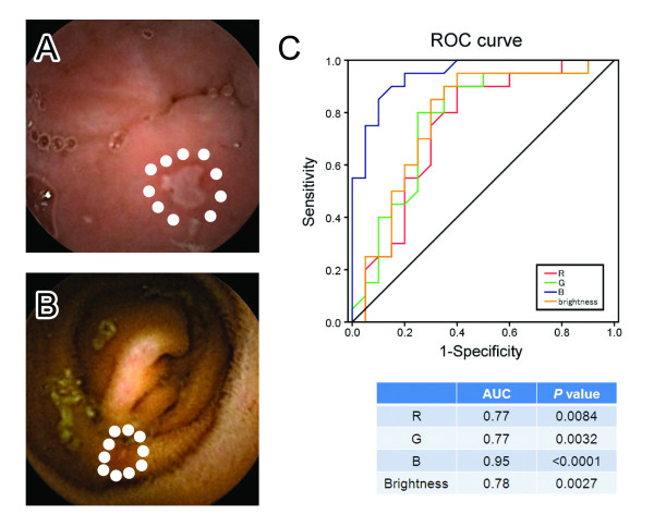
Figure 2.
Definitions of the bile-pigment-positive and bile-pigment-negative condition. CE experts selected 20 obviously bile-pigment-positive endoscopic images (A) and 20 bile-pigment-negative endoscopic images (B) that contained small bowel lesions. These images were regarded as golden standard. For each of the images, the RBG (red, blue and green) and brightness (average of ten mucosal points around the small bowel lesions, shown as white dots in this figure) values were calculated. To evaluate the most useful factor for determining whether or not bile pigments are present, a receiver operating characteristic (ROC) analysis was performed (C). The curve was obtained by calculating the sensitivity and specificity of the test at every possible cutoff point, and plotting the sensitivity against 1-specificity. The sensitivity was defined as the proportion of bile-pigment-positive images, determined from each of the color values (RBG and brightness value). The specificity was defined as the proportion of bile-pigment-negative images, also determined from the color values. As shown in Table, the results revealed that the B value was the most useful factor to determine the bile pigment condition (area under the curve (AUC): 0.95, P < 0.0001).

