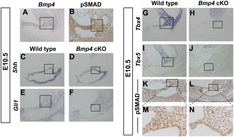
Figure 5. Defective aPCM formation of Bmp4 cKO mice.
(A) Bmp4 is expressed in the aPCM at E10.5. (B) Immunohistochemical analysis of pSMAD in the aPCM at E10.5. (C–J) Section in situ hybridization analysis with the aPCM and cloacal marker genes for wild type (C, E, G, I) and mutant embryos (D, F, H, J) at E10.5. (K–N) Immunohistochemical analysis of pSMAD in the aPCM of wild types (K) and mutant embryos (L) at E10.5. (M, N) High magnification images of square region in K and L. The squares in A–L indicate the aPCM.

