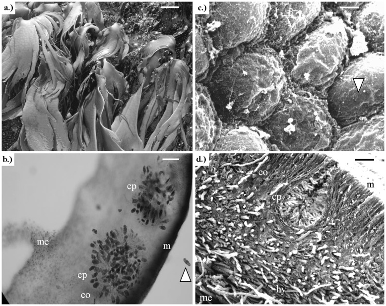
Figure 2. Durvillaea antarctica occurring at the central coast of Chile.
a) Healthy population of D. antarctica in the natural environment; b) Light microscopic microphotograph of a cross-section of a dioecious frond of D. antarctica (stained with aniline blue) showing two female conceptacles (cp) with one free oogonium (white arrow), meristoderm (m), cortical (co) and medullary (me) zones in a normal frond; c) Scanning electron microphotograph (SEM) with details of the surface of the thallus, and cells disposition in the algal surface (arrow shows one cell); d) Detail of a cross-section of a normal thallus using SEM showing early stages in conceptacle development (cp), meristoderm (m), cortical (co) and medullar (me) tissue with normal swift hyphae (hy). Scale bar: a) 10 cm; b) 100 µm; c) 2 µm; and d) 50 µm.

