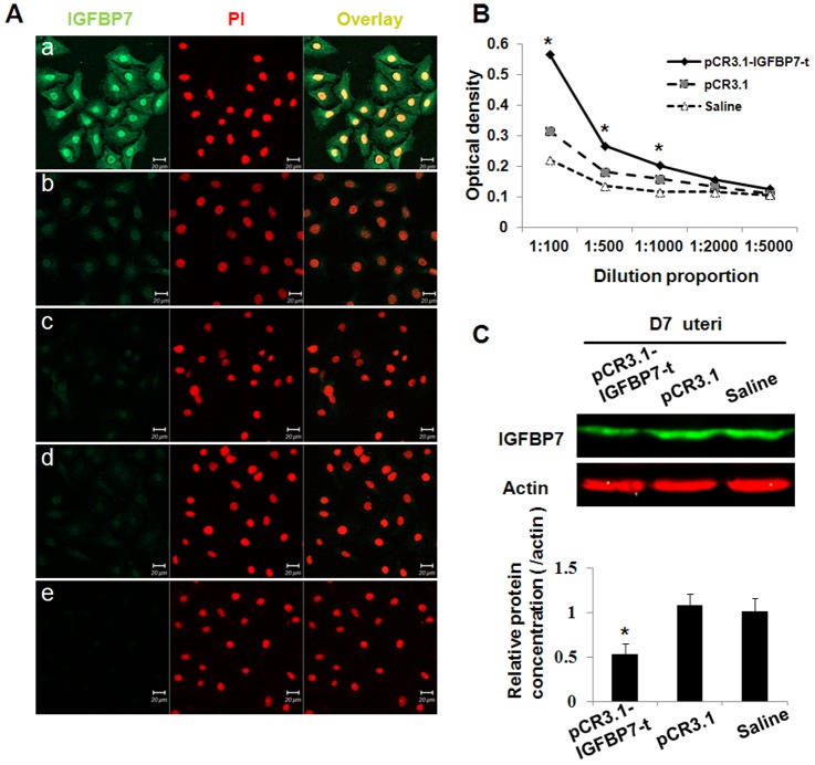
Figure 2. Detection and function of anti-IGFBP7 antibody in mice after immunizations.
A: Immunofluorescence detection of pCR3.1-IGFBP7-t expression in transfected Hela cells. Nuclei were stained with PI. a, b, c: Hela cells transfected with the pCR3.1-IGFBP7-t plasmid were incubated with antisera from mice immunized with pCR3.1-IGFBP7-t, pCR3.1 or saline respectively. d, e: Non-transfected Hela cells were incubated with antisera from mice immunized with pCR3.1-IGFBP7-t or saline respectively. B: Detection of anti-IGFBP7 antibody by Elisa assay. Antisera from mice immunized with pCR3.1-IGFBP7-t, pCR3.1 or saline were serially diluted from 1∶100 to 1∶5000. C: Western blotting analysis of IGFBP7 expression in the uteri of pregnant mice at D7. Actin served as an internal control. The relative expression of IGFBP7/actin in the uteri of the mice immunized with pCR3.1-IGFBP7-t, the pCR3.1 vector or saline were shown. IGFBP7 was significantly reduced after immunization with pCR3.1-IGFBP7-t. Bar represents 100 µm. *: P<0.05.

