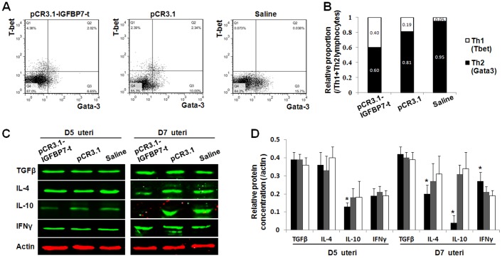
Figure 4. The analysis of uterine cytokines and T helper cells differentiation markers in mice peripheral lymphocytes after immunizations.
A: The flow cytometry detection of Tbet and Gata3 expression. The expression of Tbet was increased while the expression of Gata3 was decreased in the mice after immunization with pCR3.1-IGFBP7-t. B: the ratio of differentiated Th1 and Th2. Th1 was represented by Tbet, while Th2 was represented by Gata3. The relative proportion of Tbet (Q1) and Gata3 (Q3) were counted and shown. C: Western blotting analysis of uterine cytokines. After immunization with pCR3.1-IGFBP7-t, the expression of Th1 type cytokine IFNγ was significantly elevated and Th2 type cytokines IL-4, IL-10 were declined. No significant variation of TGFβ was observed. D: The statistics of relative expression of cytokines to actin. The black columns represent the relative expression value in mice immunized with pCR3.1-IGFBP7-t, whereas the grey columns represent the mice immunized with pCR3.1, and the white columns indicate the mice immunized with saline. *: P<0.05.

