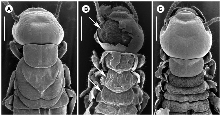
Figure 5. Scanning electron microscopy pictures.
(a) Dorsal view of the thorax of a nymph. (b) Dorsal view on a pseudergate approaching the nymphal moult, with old cuticle partially peeled off. Arrows mark compound eye (laterally on the head) and wing pads on the thorax. (c) Dorsal view of a pseudergate approaching another pseudergate instar, with old cuticle peeled off from mesothorax, metathorax and abdomen. Scale bars, 0.5 mm.

