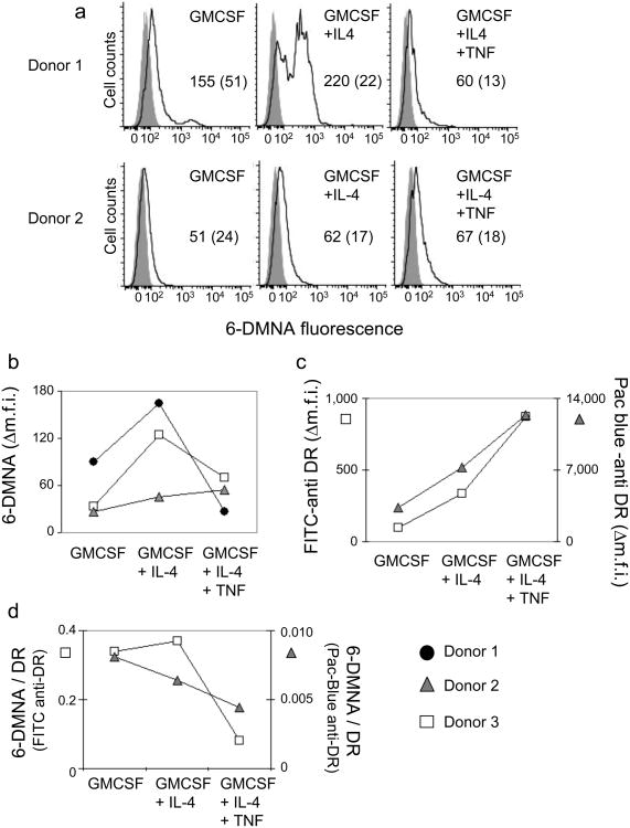
Figure 5. (6-DMNA)-peptide binds to human monocyte-derived DC in a maturation-dependent manner.
Peptide binding to monocyte derived DC subsets isolated from HLA-DR1+ donors and prepared by an in vitro differentiation protocol (see methods) was performed essentially as described for T2DR. (a) Fluorescent peptide binding to DC subsets isolated from two different donors. Solid line, (6-DMNA)-RSMA4L, shaded curve, autofluorescence. Numbers indicate the mean fluorescence intensity for peptide treated cells (with autofluorescence intensity for the same cells shown in parenthesis). (b) Δm.f.i. values for DC subsets generated from cells obtained from three different donors. (c) Expression of HLA-DR1 was measured using murine anti human HLA-DR monoclonal antibody L243 labeled with FITC or Pacific-Blue, for DC subsets from two of the donors in (b). (d) Peptide binding expressed as the ratio of 6-DMNA fluorescence to FITC-anti-DR or Pacific-Blue-anti-DR antibody fluorescence.

