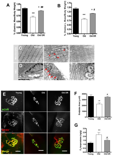
Figure 1. Calorie restriction (CR) attenuates age-related muscle atrophy.
(A) A comparison of gastrocnemius (left) and vastus lateralis (right) muscle wet-weight normalized to body weight (n=6–7). (B) Transmission electron micrographs (TEM) of interfibrillar mitochondria (C) and neuromuscular junctions (D) from gastrocnemius muscle of young, old, and old DR. Arrows denote abnormal mitochondria (n=3). Scale bar=2μm (E) A representative image of neuromuscular junction from gastrocnemius muscle. Scale bar=20μm. (F) A quantification of postsynaptic endplate area. Young mice are 7 months and old mice are 33 months old (n=6). (G) Oxidative stress is reduced by DR as measured by F2-isoprostanes as a marker of lipid peroxidation (n=6). Young vs. Old **p<0.01 and Old vs. Old DR #p<0.05. All values are represented as mean ± SEM.

