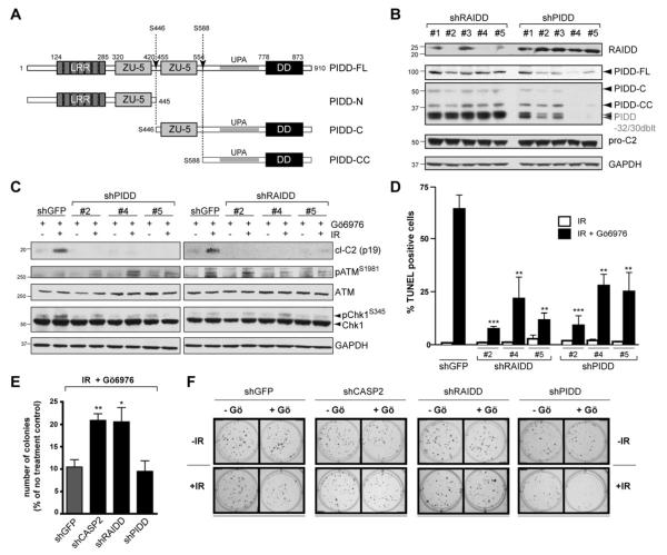
Figure 2. PIDD and RAIDD are required for CS apoptosis.
(A) Schematic diagram of PIDD maturation. Autoproteolytic cleavage sites are indicated (arrows).
(B) HeLa cells stably expressing the indicated shRNAs were analyzed by western blot. PIDD-32/30 doublet: PIDD specific bands running at ~30 kDa, presumably resulting from autoproteolysis of shorter PIDD isoforms (Cuenin et al., 2008).
(C) HeLa cells stably expressing the indicated shRNAs were treated with Gö6976 (1 μM) with or without IR (10 Gy), and harvested 24 hr post IR. Lysates were analyzed by western blot.
(D) HeLa cells stably expressing the indicated shRNAs were treated with 10 Gy IR with or without Gö6976 (1 μM) (black and white bars, respectively), and were analyzed by TUNEL staining at 48 hr post IR. Data are means +/− SEM. **p < 0.01; ***p < 0.001 two-tailed Student’s t-test.
(E) HeLa cells stably expressing the indicated shRNAs were treated with Gö6976 (1 μM) and IR (5 Gy) and colony numbers were recorded 14 days post IR. Data are means +/− SEM. *p < 0.05; **p < 0.01 two-tailed Student’s t-test.
(F) Representative images of a clonogenic assay. HeLa cells stably expressing the indicated shRNAs were treated with or without Gö6976 (1 μM) or IR (2 Gy) and stained with crystal violet 14 days after IR.

