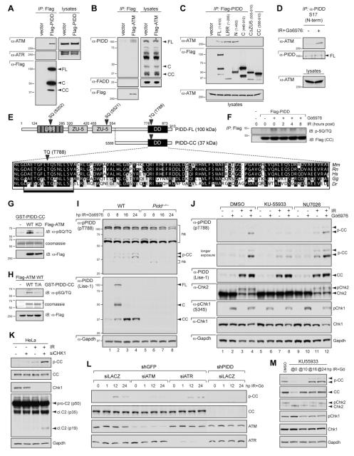
Figure 4. ATM phosphorylates PIDD on T788 during CS apoptosis.
(A) HeLa cells transfected with Flag-tagged PIDD were lysed, immunoprecipitated with anti-Flag antibodies and analyzed by western blot.
(B) HeLa cells transfected with Flag-ATM were lysed, immunoprecipitated with anti-Flag antibodies and analyzed by western blot.
(C) HeLa cells transfected with the indicated Flag-tagged PIDD deletion constructs were lysed 24 hr post transfection, immunoprecipitated with anti-Flag antibodies and analyzed by western blot.
(D) HeLa cells either untreated or treated with IR (10 Gy) and Gö6976 (1 μM) were harvested 24 hr post IR. Nuclear extracts were immunoprecipated with an anti-PIDD antibody targeted to the PIDD N-terminus. Immunoprecipitates were analyzed by western blot.
(E) Diagram of PIDD-FL highlighting three candidate ATM phosphorylation sites (bold arrowheads). Blow up shows a clustal alignment of the DD amino acid sequences of PIDD proteins from the indicated species. Mm, Mus musculus; Rn, Rattus norvergicus; Hs, Homo sapiens; Gg, Gallus gallus (chicken); Dr, Danio rerio (zebrafish). The predicted full ATM target sequence (box) and the target TQ motif (bold arrowhead) are indicated.
(F) HeLa cells transfected with empty vector or C-terminally Flag-tagged PIDD were treated with IR (10 Gy) with or without Gö6976 (1 μM) and harvested at the indicated time points after IR. Protein extracts were immunoprecipitated with anti-Flag antibodies and analyzed by western blot.
(G) Recombinant GST-PIDD-CC proteins were incubated with wild-type (WT) or kinase-dead (KD) Flag-ATM for an in vitro kinase assay (IVK). Reactions were analyzed by coomassie staining and western blot.
(H) Recombinant wild-type (WT) or T788A mutant (T/A) GST-PIDD-CC proteins were incubated with Flag-ATMWT for an IVK. Reactions were analyzed by coomassie staining and western blot. Residual pSQ/TQ immunoreactivity in the T/A sample suggests additional specific or non-specific phosphorylation events occurring in the reaction.
(I) SV40 MEFs of indicated genotypes were treated with IR (10 Gy) and Gö6976 (1 μM) and harvested at the indicated time points after IR. Extracts were analyzed by western blot. p-CC, phospho-PIDD-CC; upper arrowhead marks p-CC harboring an additional post-translational modification (see text). ns, non-specific bands.
(J) SV40 WT MEFs treated with or without IR (10 Gy) or Gö6976 (1 μM) were additionally treated with ATM inhibitor KU55933 or DNA-PKcs inhibitor NU7026 (10 μM each) at 8 hpIR. Cells were harvested at 24 hpIR and analyzed by western blot.
(K) HeLa cells transfected with CHK1 siRNA (+) or LACZ siRNA (-) were treated with or without IR (10 Gy) or Gö6976 (1 μM) at 16 hr post-transfection. Cells were harvested at 16 hpIR and analyzed by western blot.
(L) HeLa cells stably expressing the indicated shRNAs were transfected with the indicated siRNAs and treated with or without Gö6976 (1 μM) or IR (10 Gy) 36 hr post-transfection. Cells were harvested at the indicated time points after IR and analyzed by western blot.
(M) SV40 WT MEFs treated with IR (10 Gy) and Gö6976 (1 μM) were exposed to ATM inhibitor KU55933 added at (‘@’) the indicated time points after stimulus. Cells were harvested at 24 hr after IR+Gö6976 treatment and analyzed by western blot.

