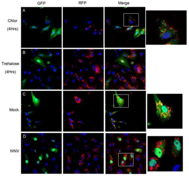
Figure 5. WNV infection results in acidification of autophagosomes.
Vero cells were transfected with a plasmid expressing GFP-RFP-LC3 and treated with A) chloroquine (50μM) or B) trehalose (10mM) as assay controls. Cells were fixed and harvested at 4 hours post-treatment for fluorescent imaging. Chloroquine treated cells display continued GFP signal but trehalose activation of autophagy results in a shift to RFP signal due to autophagosome acidification. GFP-RFP-LC3 transfected cells were inoculated with C) mock or D) WNV (MOI 3) and cells were harvested at 24 hours post-infection. Mock infected cells display persistent GFP signal (green and yellow) while WNV infected cells exhibit a shift to RFP (red) signal. Images shown at 600x original magnification with boxes representing digitally magnified cells in the same row (1000x). Images representative of 3 replicate experiments and all nuclei were stained blue with DAPI.

