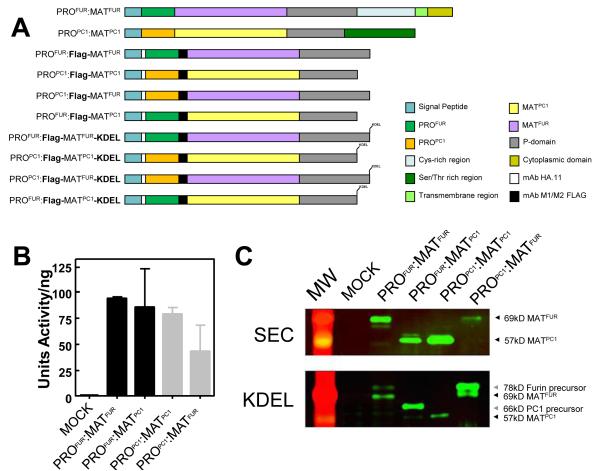
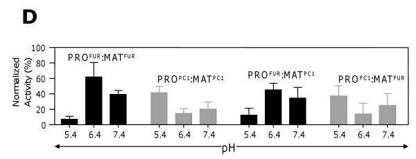
Fig. 3. Transferring propeptides between eukaryotic subtilases reassigns their optimum pH for activation.
(A) Schematic of constructs for Furin and PC1. (B) Normalized protease activity assayed in conditioned media (CM) from Cos-7 cells transfected with 2 μg of DNA (C) Western blot analysis of CM from cells expressing secreted reporter constructs (top panel; SEC), and ER fractions from cells expressing KDEL-tagged reporters probed using mAB-M2. Molecular weight of each species is indicated by the arrowheads; Unprocessed furin, 78kD; Processed furin, 69kD; Unprocessed PC1, 66kD; Processed PC1 57kD. (D) pH-dependent activation of KDEL-tagged reporters measured after incubating ER membrane fractions at designated pH29. Maximal activity was estimated by trypsinizing membrane fractions for 1 hr and inhibiting trypsin by soybean trypsin inhibitor prior to the protease assay.


