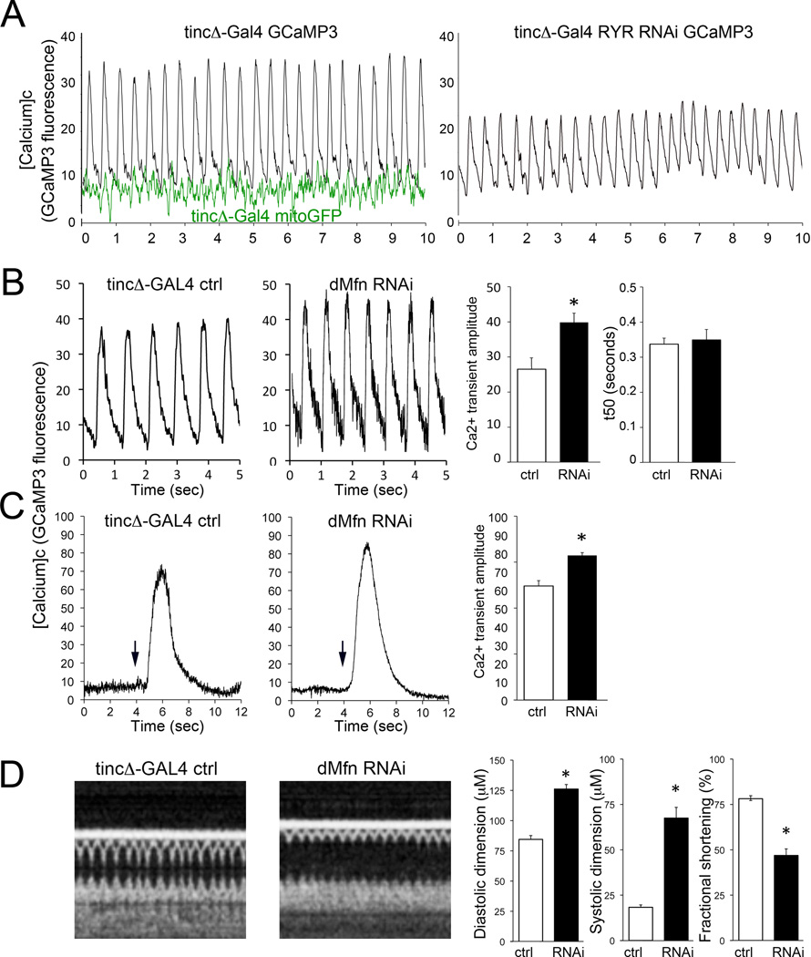
Figure 1. SR Ca2+ handling is altered by dMfn/MARF suppression in Drosophila heart tubes.
A. (left) Representative Ca2+ transients monitored by fluorescence of GCaMP3.0 expressed specifically in cardiac myocytes of spontaneously contracting Drosophila heart tubes in situ. Black tracings are tincΔ-Gal4-driven GCaMP3 (controls); green tracings are tincΔ-Gal4 driven mitoGFP (which is Ca2+-insensitive), demonstrating minimal effects of heart tube contraction on fluorescence signals. (right) Representative tracing of RyR-deficient Drosophila heart tube, showing decreased amplitude and delayed normalization of Ca2+ transient, which is typical of heart failure. B. Ca2+ transients of control (ctrl; tincΔ-Gal4) and mitofusin/MARF deficient (dMfn RNAi) expressing RNAi for dMfn. Representative tracings are shown to the left and group data from n=11 individual flies are to the right. C. SR Ca2+ content measured as caffeine-stimulated cardiomyocyte Ca2+ release in heart tubes of dMfn deficient (RNAi) and ctrl flies. Representative tracings are shown to the left with arrows marking the time of caffeine (10 mM) addition; group data from n=5 or 6 individual flies are to the right. *p<0.05 vs ctrl. D. Heart tube dimensions and contractile function in control and dMfn RNAi Drosophila, assessed by optical coherence tomography (OCT). Representative b-mode OCT scans are shown on the left, with group data from the same set of flies that underwent Ca2+ measurements shown to the right. *p<0.05 vs ctrl.

