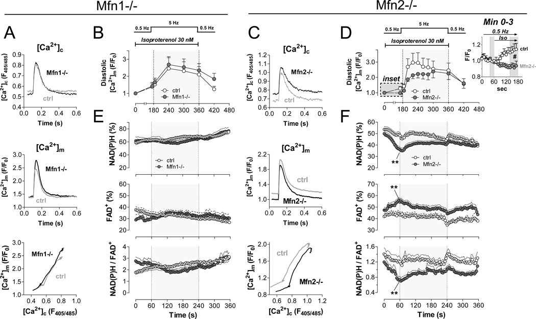
Figure 5. Impaired mitochondrial Ca2+ accumulation and bioenergetic feedback response in Mfn2-deficient myocytes.
Experiments were performed on intact cardiac myocytes with acute isoproterenol and pacing stress (see Supplemental Figure 4); Mfn1-KO on left; Mfn2-KO on right. A. and C. Averaged original traces of [Ca2+]c (top) and [Ca2+]m transients (middle) in WT and Mfn-KO myocytes after isoproterenol (30 nM) for 1 min at 0.5 Hz. Bottom panels show dynamic changes of [Ca2+]m plotted against [Ca2+]c in the same cell in the presence of isoproterenol for 1 min at 0.5 Hz (Mfn1: n= 4 control, n=7 KO; Mfn2: n=14 control, n=12 KO). B. and D. Time-dependent changes in diastolic [Ca2+]m with pacing and isoproterenol stress. Inset in D shows change of diastolic [Ca2+]m in the first 2 minutes after application of isoproterenol. E. and F. Autofluorescence of NAD(P)H (top), FAD (middle) and the ratio of NAD(P)H/FAD (bottom; Mfn1-KO, n=17; control, n=7; Mfn2-KO, n=24; control, n=15). *p<0.05 and **p<0.01 WT vs. KO, respectively (ANOVA for repeated measures).

