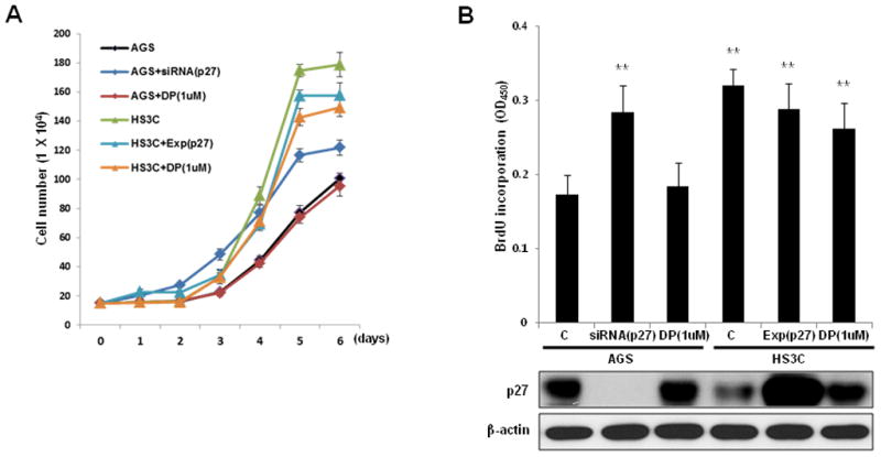
Fig. 6.

Effects of p27 on gastric epithelial cell proliferation. AGS cells were transfected with a control siRNA, p27 siRNA, or treated with 1 uM DPDPE (DP). HS3C cells were transfected with a control plasmid, p27 expression plasmid, or treated with 1 uM DP. The monolayer growth rates of cells were determined with cell counting (A) and a BrdU incorporation assay (B). The cell number was counted on the indicated day, and the BrdU incorporation assays were conducted at 48 h after cellular transfections. The levels of cellular p27 in association with BrdU incorporation are shown at the bottom of (B). The data represent the mean ± SD of 3 independent experiments with **P < 0.01 versus the corresponding control.
