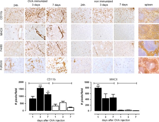
Fig. 3.

Macrophage and microglia activation in the brain after intracerebral injection of OVA. OVA-immunized mice or control non-immunized mice received a unilateral injection of OVA into the striatum and were assessed for presence of macrophage and microglia activation. Data shows CD11b, MHCII, F4/80 and FcγRII/III immunoreactivity after 24 h, 3 days or 7 days in OVA-immunized mice or non-immunized mice. As a positive control for these well-characterized antibodies spleen tissue from a naïve mouse was used. The number of DAB-positive pixels (cells and their processes) in OVA-immunized (black bars) and non-immunized mice (white bars) was quantified as described in “Materials and methods”. Statistical analysis: One-way ANOVA, Dunnett’s post-test *p < 0.05. Data represents the mean of n = 3–5 per treatment and time point. The experiment was performed twice independently with comparable results
