
Fig. 2.
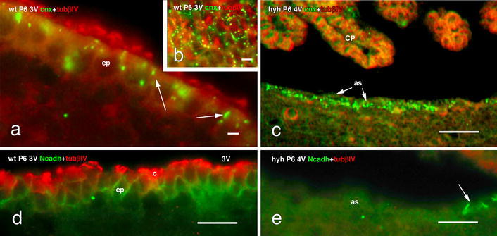
Expression of junction proteins in the cell lining the floor of the fourth ventricle of wt and hyh mice at P6. a–c Double immunofluorescence for tubulin βIV (tubβIV, red) and connexin 43 (cnx, green). a The multiciliated ependyma (ep) in wt mice present cnx+ spots localized preferentially at the apical cell pole of ependymal cells (arrows). b Tangential section through the apical cell poles of the ependyma showing the distribution of cnx as dots in the lateral plasma membrane. c The astrocyte layer (as) lining the denuded ventricle in the hyh mouse expresses cnx (arrow) but not tubβIV. d, e Double immunofluorescence for tubβIV (red) and N-cadherin (Ncadh, green). d The ependyma in wt mice shows the belt-like distribution of Ncadh. e The astrocytes lining the denuded ventricles do not express Ncadh. Arrow points to a few ependymal cells remaining in situ and expressing Ncadh. 3V third ventricle, 4V fourth ventricle, c cilia, CP choroid plexus. Scale bars a 6 μm, b 8 μm, c 40 μm, d 12 μm, e 12 μm