
Fig. 3.
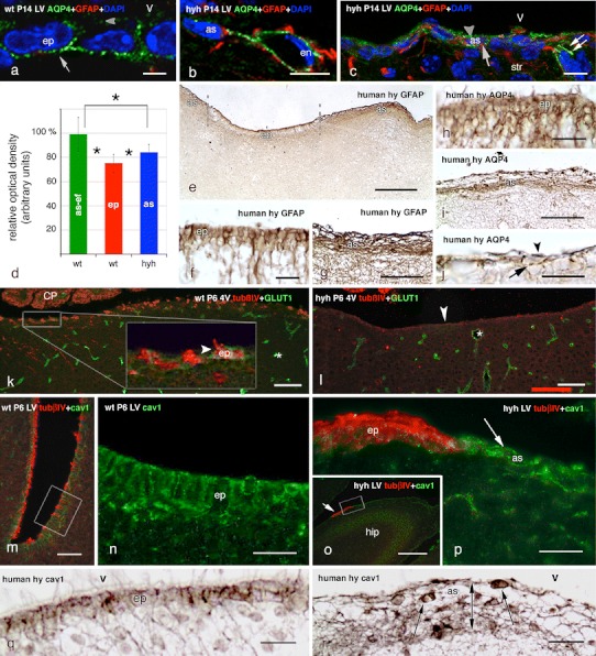
Expression of aquaporin 4, the glucose transporter 1 (GLUT1) and caveolin-1 in the ependyma of wt mice and in the astrocyte layer that covers the ependymal-denuded surface in hyh mice and a human hydrocephalic foetus. a Immunolabelling for aquaporin 4 (AQP4, green) in the ependyma (ep) of the lateral ventricle (LV) of a wt mouse; DAPI counterstaining (blue). The water channel is mainly located in the baso-lateral plasma membrane domain (arrow), but there is also a weak reaction at the apical domain (arrowhead). b, c Latero-medial wall of a hyh mouse with double immunofluorescence for GFAP (red), AQP4 (green) and DAPI counterstaining (blue). AQP4 immunoreaction is present in a perivascular astrocyte (as) and its endfeet surrounding endothelial (en) cells (b). Reactive astrocytes present AQP4 at the apical (arrowhead) and basal (arrow) cytoplasm and in their cell processes (double arrow) (c). d Optical density of the immunoreaction for AQP4 was recorded at (1) the ependyma (ep) of the latero-medial wall of the lateral ventricles of wt mice; (2) the cell layer of reactive astrocytes lining denuded areas of the latero-medial wall of the lateral ventricles of hyh mice (as); (3) the perivascular endfeet of the astrocytes (as-ef) of wt mice. Data represent the mean and standard deviation from four wt and four hyh mice (3–4 sections each mouse; four neighbour areas from each section). Data are expressed as relative percentage of the values obtained in each section where the mean of as-ef in wt mice was considered to be 100 %. *Correlation analysis showed significant differences (p < 0.001, Student’s t test). e–j Lateral ventricle of a 40-week-old human hydrocephalic foetus. Immunostaining for GFAP (e–g) and AQP4 (h–j). e Low power view showing ependyma not yet disrupted (ep) and denuded areas lined by a layer of GFAP+ astrocytes (as). These two regions are shown at higher magnification in f and g. The ependyma (h) and the astrocyte layer (i, j) express AQP4. k, l Fourth ventricle (4V) with double immunofluorescence for tubulin βIV (tubβIV, red) and GLUT1 (green). Endothelial cells (asterisks) are reactive to GLUT1. k The inset shows a weak reaction for GLUT1 in the ependyma lining the floor of the fourth ventricle of a P6 wt mouse. l The astrocyte layer lining the denuded floor of the fourth ventricle of hyh mice does not express GLUT1. m Lateral ventricle of a wt P6 mouse. Double immunofluorescence for tubβIV (red) and caveolin-1 (cav1, green). The framed area is shown in n using only the cav1 channel. n Detailed view of area framed in m, showing the strong expression of cav1 in the multiciliated ependyma of the lateral ventricle. o Wall of the lateral ventricle close to the hippocampus (hip) of a P6 hyh mouse. Double immunofluorescence for tubβIV (red) and cav1 (green). The walls are denuded with the exception of a tubβIV+ resistant ependymal patch (arrow). The area framed is shown in p. p Astrocytes lining the denuded areas of the lateral ventricle strongly express cav1 (arrow). The patch of ependyma is strongly labelled for tubβIV (red), whereas the astrocyte layer is not. q, r Lateral ventricle of a 40-week-old hydrocephalic human foetus. Section adjacent to that shown in f and g, immunostained for cav1. The cell body of ependymal cells contains immunoreactive granules (q). The cell body (arrows) and processes of the astrocytes forming the thick cell layer lining denuded areas (double-ended arrow) are immunoreactive (r). CP choroid plexus, V ventricle lumen. Scale bars a 5 μm, b, c 10 μm, e 200 μm, f, h, j 20 μm, g 50 μm, i 50 μm, k, o 100 μm, l 80 μm, m 40 μm, n, p 20 μm