
Fig. 4.
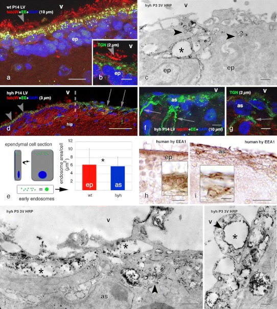
Endocytosis of HRP injected in vivo into the lateral ventricle of wt and hyh mice, and location of endocytic vesicles, early endosomes and the Golgi apparatus. Lateral ventricle of wt (a, b) and hyh (d, f, g) mice, at P14. Confocal laser microscopy of immunolabelling for the EEA1 antigen of early endosomes (EE, green in a, d, f), the trans-Golgi network (TGN, green in b, g) and tubulin βIV (tubβIV, red). DAPI nuclear staining (blue). Z-projections comprising confocal planes for different thicknesses (between brackets). Numerous EE are present in the apical pole (arrowheads in a, d) of the ependymal cells (ep) in wt and hyh mice. The broken line in d shows the border between a patch of intact ependyma (arrowheads) and the astrocyte layer covering an ependyma-denuded surface (arrows) in the hippocampus (hip). The TGN in ependymal cells is located juxtanuclear (arrowhead in b). In the astrocytes (as) covering the ependyma-denuded surface of hyh mice (arrows in d), abundant EE and TGN are present in the cell bodies and processes (arrows in f and g). e Total area occupied by EEA1-reactive EE per ependymal cell of wt mice and per reactive astrocyte (as) of hyh mice in confocal laser cuts of frozen sections (explanation on the left side of the figure). Data represent the mean and standard deviation obtained from sections corresponding to four wt and four hyh mice (4 sections each mouse). *Correlation analysis did not show a significant difference (p = 0.695, Student’s t test). c, j, k Ultrastructural detection in the third ventricle (3V) wall of HRP injected into a lateral ventricle at P3 in wt mice (c) and hyh mice (h, i). In the apical pole of ependymal cells and in astrocytes, HRP is located within endocytic vesicles (arrowheads) and large EE (asterisks). h, i Lateral ventricle of a 40-week-old human hydrocephalic foetus. Section adjacent to that shown in Fig. 3f, g, immunostained for EEA1. The cell body of ependymal cells (ep) contains immunoreactive granules (h). The cell body and processes of the astrocytes forming the thick cells layer lining denuded areas are immunoreactive (i). LV lateral ventricle, str striatum, V ventricle lumen. Scale bars a 10 μm, b, g 5 μm, c, j 400 nm, d 50 μm, f 7 μm, h, i 20 μm; Insets in h, i 10 μm, k 200 nm