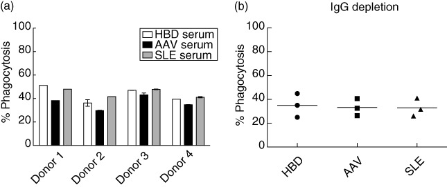
Fig. 4.

Phagocytosis in the presence of serum. (a) Four healthy blood donors (HBD) monocyte-derived macrophages (MØ) donors were incubated with CellTracker™ Green-stained apoptotic Jurkat cells at a MØ : Jurkat ratio of 1:2 in the presence of 10% pooled serum from HBDs, anti-neutrophil cytoplasmic antibody (ANCA)-associated vasculitis (AAV) or systemic lupus erythematosus (SLE) patients with eight donors in each pool. A decrease in phagocytosis was seen when adding AAV serum compared to HBD- and SLE serum, although this did not reach significance. This graph represents one experiment for each donor, but with duplicate samples for all except MØ donor 1. (b) Immunoglobulin (Ig)G depletion of serum from the same serum pools as in (a). This experiment was performed as in (a), but with three HBD MØ donors. Percentage phagocytosis is measured as % CellTracker™ Green-stained CD206-positive MØ. Statistics were calculated using the Mann–Whitney test and error bar represents standard deviation in cases where duplicate samples exist.
