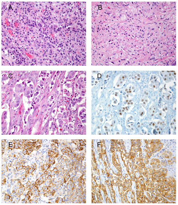
Figure 1.
A) Infiltrating poorly differentiated UC.
B) Gleason score 10 prostatic adenocarcinoma.
C) Invasive poorly differentiated UC.
D) GATA3with both moderate and strong nuclear staining (same case shown in 1C).
E) UC showing diffuse and strong staining with THROMBO.
F) UC showing diffuse and strong staining with Uroplakin III.

