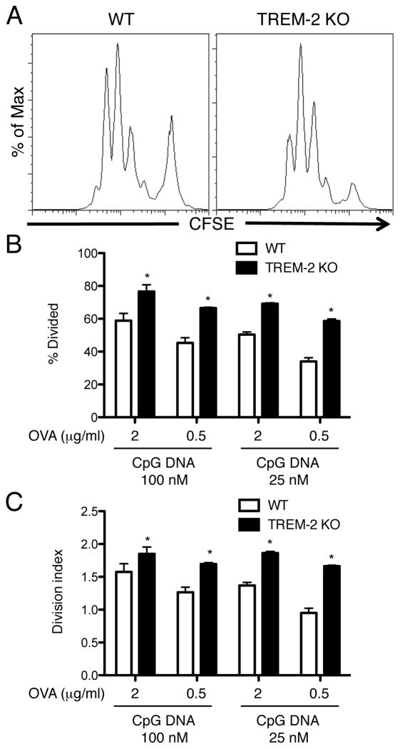
Figure 6. Enhanced antigen-presentation activity in TREM-2-deficient DCs.
(A) CD11c-purified BMDCs from WT and TREM-2-deficient mice were co-cultured with CFSE-labeled CD4+ OT-II T cells in the presence of OVA, CpG DNA and GM-CSF for 72 h. After co-culture, CFSE dilution of CD4+ OT-II T cells was detected by flow cytometry. (B) The percentage of divided and (C) division index of CD4+ T cells were calculated by Flowjo software. Data are represented as mean +/− SD of triplicate wells. *p<0.05 versus WT, as determined by an unpaired two-tailed students t-test. These data are representative of two independent experiments.

