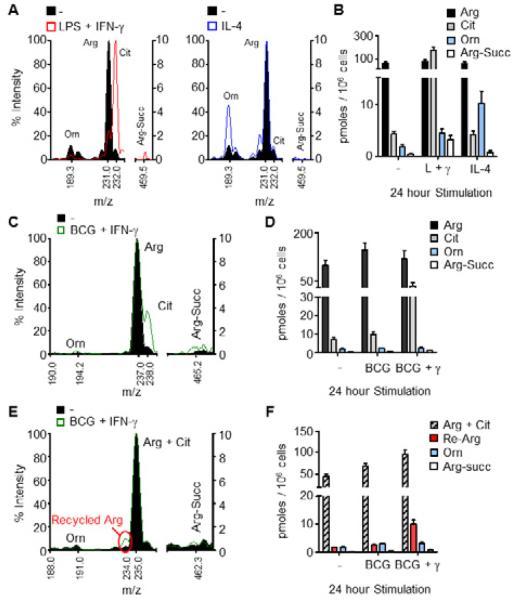
Figure 2. Detection of arginine metabolites by mass spectrometry (MS).
(A and B) BMDMs were untreated (-) or stimulated with LPS + IFN-γ or IL-4 for 24 hours, and cell lysates were analyzed by MS for arginine (Arg), citrulline (Cit), argininosuccinate (Arg-Succ), and ornithine (Orn). Sample mass spectra are shown (A) with the corresponding concentrations of indicated metabolites (B).
(C-F) BMDMs were infected with BCG with or without IFN-γ in the presence of heavy 13C-arginine (C, D) or 15N-arginine (E, F) for 24 hours, and cell lysates were analyzed by MS as above. The right y-axis was used for MS analysis of argininosuccinate (C, E). Data are representative of at least 3 experiments (A, B) or one experiment (C-F) with BMDMs pooled from at least 4 mice. Recycled arginine (Re-Arg).
Error bars, SEM.
See also Figure S2.

