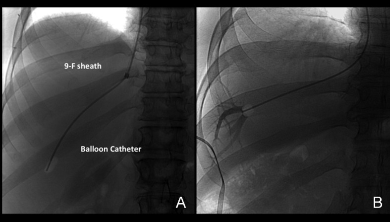
Figure 5.
Wedge position using an occlusion balloon. (A) Anteroposterior (AP) spot film demonstrates the occlusion-balloon catheter inflated with a small amount of air. The catheter is positioned in a peripheral location within the hepatic vein. (B) AP spot film obtained during injection of a small amount of contrast with the occlusion balloon inflated with a small amount of air. Note opacification of the peripheral hepatic vein branches. No contrast leak is noted around the balloon, indicating a good wedge position.

