
Figure 4.
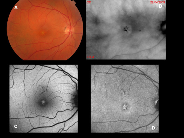
A: Color photograph of Adult-onset foveomacular vitelliform dystrophy. B: Retromode imaging shows focally risen lesions with darker borders. C: BL-AF reveals focally increased signal corresponding to the accumulation of abnormal material. D: NIR-AF displays a more intensely hyper-autofluorescent lesion.