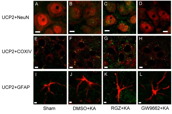
Figure 5.
Laser scanning confocal microscopic images of the right CA3b subregion of hippocampus showing cells that were immunoreactive to a neuronal marker, a mitochondrial protein marker, or a marker for astrocytes, and additionally stained for uncoupling protein (UCP)-2. Scanning was performed 24 h after microinjection of 0.5 nmol kainic acid (KA) or PBS into the left hippocampal CA3 subfield in animals that received pretreatment with application into the bilateral CA3 subfield of 3% dimethyl sulfoxide (DMSO), 6 nmol rosiglitazone (RGZ) or 500 ng GW9662. (A-D) Neuronal marker, NeuN (red fluorescence). (E-H) Mitochondrial protein marker, cytochrome c oxidase subunit IV (COX IV) (red fluorescence). (I-L) Marker for astrocytes, glial fibrillary acidic protein (GFAP) (red fluorescence). Cells were additionally stained for UCP2 (green fluorescence). Note that double-labeled neurons, mitochondria or astrocytes displayed yellow fluorescence. These results are typical of three animals from each experimental group. Scale bar, 5 μm in 5A-H; 2 μm in 5I-L.

